
Anaplastic Thyroid Cancer (ATC)
Anaplastic thyroid cancer is the least common of the thyroid cancers accounting for 0.5-1.5% of the total. Its peak incidence is in patients over 60 years of age, and is most common in areas of endemic goitre where there is chronic iodine deficiency. The incidence of ATC has steadily decreased over the past few decades, although the reason for this decline is not completely understood, and several factors may be involved.
There are three main types of anaplastic cancer, each with a differing likelihood of being able to be resected. First there is the previously unsuspected rapidly growing mass (can rarely resect), second is the PTC or FTC transforming into ATC (resection may be possible), and third is the ATC arising in a goitre (8% of ATC), usually found incidentally, occuring in an endemic goitrous region (resection often already done, but if not, worth trying).
The cancer may arise in pre-existing follicular tumours. The progression of PTC or FTC to ATC has been well documented at a clinical and molecular level with the loss of the p53 tumour suppressor gene. In one study up to 76% of ATC had previous or concurrent thyroid disorders, with 47% related to well differentiated thyroid cancer.
Presentation
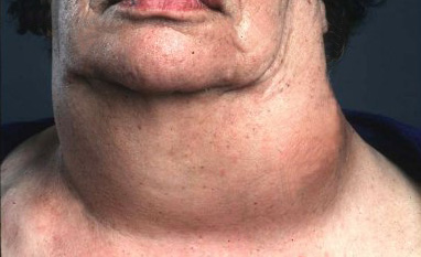 Fig.1: Large left anaplastic cancer of thyroidATC is a very aggressive tumour with a poor prognosis, being one of the most lethal cancers in humans. Even with modern treatments, less than 10% of patients survive more than 3 years.
Typically there is a history of a long-standing goitre that suddenly increases in size, with the onset of increasing local compressive symptoms (Fig. 1).
As the tumour invades the trachea and the oesophagus it may present with stridor (noisy breathing) and dysphagia (difficulty swallowing). A change in the voice, pain and neck tenderness are common, and the mass tends to be hard and fixed to other structures.
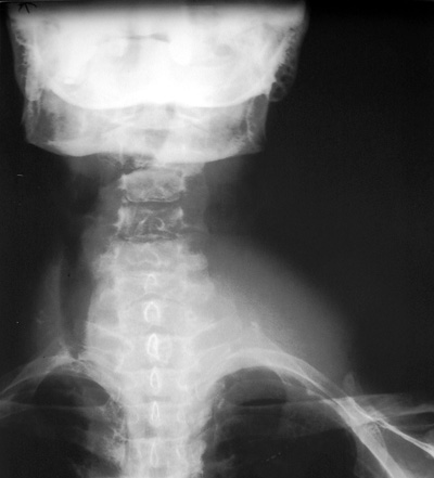 Fig.2: Xray showing soft tissue tumour on left of the neck, and gross tracheal displacement to right
At the time of diagnosis local lymph node metastases are seen in 40% and vocal cord paralysis in up to 30% of patients with ATC. Over 70% of ATC patients have direct invasion of surrounding tissues, such as fat, trachea, muscle, oesophagus, and larynx.
Systemic metastases occur in up to 75% of patients, with lung being the most common site (80%), followed by bone (6% to 15%) and brain (5% to 13%).
Investigation with FNA cytology and thyroid imaging will make the diagnosis. CXR and CT scan will often show tracheal narrowing or displacement (Figs. 2 & 3).
 Fig.3: CT Scan of large anaplastic cancer pushing the trachea and oesophagus to the rightTreatment
There is no definitively effective therapy for ATC. Age <60 years, a smaller (<7cm) cancer confined to the thyroid, without distant metastases, and treated with combination surgery/radiotherapy treatment seem to be the only factors that might increase survival rates.
Although it accounts for less than 1% of all thyroid cancer cases, it accounts for 14 to 39% of all cancer deaths.
In the majority of cases surgery is limited to an open biopsy to exclude lymphoma (a tumour with a better prognosis than anaplastic cancer), although core biopsy may be enough. Sometimes removal of the isthmus is necessary to free the windpipe from the tumour.
Rarely the anaplastic tumour is confined within the thyroid, the patient has a normal voice and the trachea, carotid and oesophagus are not involved. In these rare circumstances an aggressive approach is justified and a total thyroidectomy possible, followed by chemoradiation. If speech or swallowing is affected by the tumour, total thyroidectomy is impossible and resection of the thyroid should not be considered.
There is no role for radioiodine as ATC does not take it up, so the mainstay of treatment in extensive ATC is external beam radiotherapy. Unfortunately initial shrinkage in the tumour is short lived and regrowth inevitable. Chemotherapy with doxorubicin, 5-fluorouracil and cisplatin, or paclitaxel is usually combined with external-beam radiotherapy, although the results are poor whether single or multiple drug regimes are used. There is some evidence that the tyrosine-kinase inhibitor, sorafenib, may help.